Referral
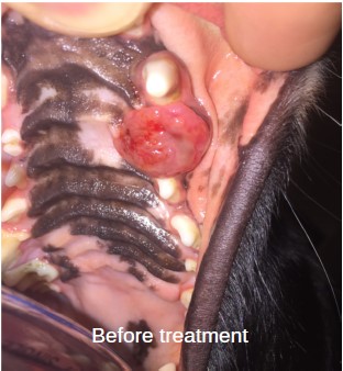
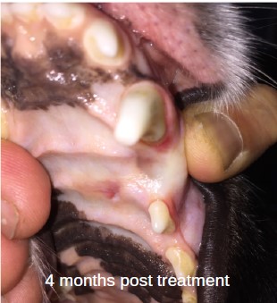
A 2-year-old female border collie was referred to the oncology team to discuss radiation therapy for a left maxillary mass with intranasal extension crossing the midline of the hard palate.
Patient History
The patient was initially evaluated after bleeding from the mouth was noted after playing with a rope. On oral exam a large friable mass was seen. A biopsy of the mass was obtained by the family vet which was consistent with an undifferentiated carcinoma, and she was referred to the oncology team in Canberra for further evaluation.
Diagnostics
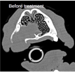
A skull CT was done which showed a large contrast enhancing mass involving the maxilla bone and left dental arcade from the maxillary canine rostrally and extending caudally toward the premolars. In addition extension of the lesion into the nasal passage was noted and it crossed over the midline of the hard palate. Based on the extent of the tumour, the patient was not a good candidate for surgery, and she was referred to the oncology team in Sydney to discuss radiation therapy options. A CT simulation for radiation planning was done on 12/6/2017.
Outcome
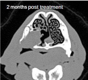
In September 2017 , 12 weeks post radiation a CT scan showed a 65% reduction of the original volume of the mass. The patient moved overseas and passed away 4 years and 2 months after completion of treatment.
Stereotactic radiation therapy (SRT) 3 day course of treatment to the left maxilla and intranasal extension

SRT was prescribed to the left maxilla as a non-surgical alternative. Treatment was delivered via volumetric arc therapy (VMAT)

30Gy in 3 fractions to the left maxilla mass and intranasal extension

VMAT fields are used to provide excellent treatment preserving healthy tissue and critical surrounding structures.

Treatment planning utilized CT imaging with daily treatment QA achieved with cone beam CT scans.

15 minute treatment time with dailyimage guided radiation therapy (IGRT) used to verify the accuracy and precision of treatment.
