Referral
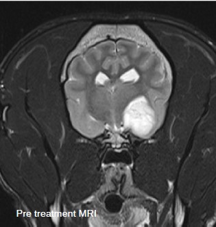
A 7-year-old male French Bulldog was referred to discuss radiation therapy for a right piriform lobe tumour suspected to be a glioma based on imaging characteristics.
Patient History
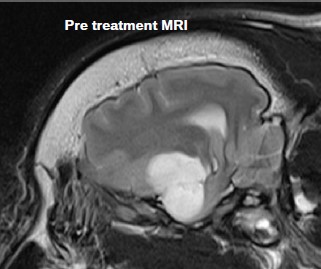
The patient reportedly had his first seizure in December 2016 after a rigorous walk. He was started on 45mg phenobarbital twice daily at that time and di reasonably well until a recent onset of further seizure activity in June 2017. This prompted further evaluation at a referral hospital where an MRI diagnosed an approximately 2 x 1.5cm diameter intra-axial mass in the right piriform lobe.
Observations
Given the lesion location surgery was not a viable option and the patient was referred to the oncology team for evaluation for radiation therapy. CT simulation for stereotactic radiation therapy was done on 26/6/2017. These images were fused with the MRI for radiation planning.
Outcome
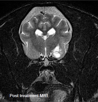
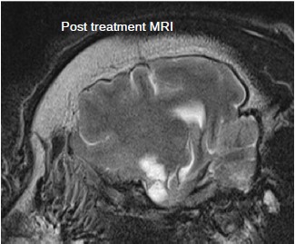
The patient tolerated the treatment very well with no acute adverse effects. A follow up MRI done 3 months later showed significant mass reduction. In July 2018 (13 months post SRT) the patient had several additional seizures. A repeat MRI at this time showed the mass remained markedly reduced in size. His anti-epileptic medications were adjusted, and the seizures were controlled again. At 14 months post SRT the patient was clinically stable with a normal neurological examination. The patient passed away 21 months after diagnosis.
Stereotactic radiation therapy (SRT) 3 day course of treatment to the right piriform lobe

SRT was prescribed to a mass in the right piriform lobe as a non-surgical alternative.
24Gy in 3 fractions to the right piriform lobe Glioma

VMAT fields are used to provide excellent treatment, preserving healthy tissue and critical surrounding structures.

Treatment was planned via CT planning scan.

15 minute treatment time with daily image guided radiation therapy (IGRT) used to verify the accuracy and precision of treatment.
